Our next researchers in the #BearonTour series are Hector Basevi and Joao Correia from The Centre of Membrane Proteins and Receptors (COMPARE). This is a unique cross-University Research Centre set up between the Universities of Birmingham and Nottingham.
COMPARE’s research focuses on better understanding the mechanisms responsible for cell communication and the role they play in physiology and disease, aiming to develop innovative therapies for common conditions such as cardiovascular disease, diabetes or cancer.
These efforts are supported by dedicated state-of-the-art facilities, including an Advanced Imaging Facility. The facility is open to internal and external researchers in academia and industry, and provides access to cutting-edge bioimaging technologies covering multiple modalities across scales, from single-molecules to whole organisms, enabling the investigation of intricate biological and biomedical questions.
In addition, COMPARE can also offer access to high performance computing resources and sophisticated image analysis software to analyse the complex data produced by COMPARE microscopes.
Research portal (Hector) Research portal (Joao)
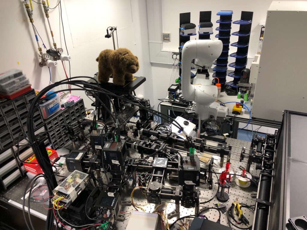
COMPARE’s latest high-end imaging system (under development and undergoing final testing) is a custom 5-camera OPLSM (Oblique Plane Light Sheet): this is a fully automated, remotely accessible system combining high content imaging with light sheet microscopy. COMPARE owns high performance computing (HPC) resources managed by BEAR which will run some of the microscope’s AI systems. These AI systems analyse raw data produced by the imaging system to automatically find cells of interest and decide in realtime where and when to image. These HPC resources are also available for other AI and data analysis use cases.
Hector’s interview
Q1 – Tell me about your research
I’m working as a Research Fellow within COMPARE on machine learning-based generative AI for modelling cell behaviour over time and how that behaviour influences the content of 3D microscopy videos. Modern high-throughput microscopes can generate a huge amount of data, imaging thousands of cells in 3D at high resolution and framerate and recording the spatiotemporal distributions of glowing molecules, which have been labelled by the researcher via fluorescent markers. Analysing how these molecules move over time and interact with parts of the cell and each other can shed light on communication pathways inside the cell and cellular behaviour. However, standard analysis techniques can struggle with the volume of data and can only perform a small set of tasks such as detecting and segmenting cell components.
Conversely, for AI the bigger the datasets the better, and by designing the structure of the AI system appropriately, it may be possible to implicitly learn something about the underlying biochemical processes influencing behaviour. I’m building systems which model the image formation process in the microscope and incorporate neural networks to represent the individual organelles that form the cell and model how they interact and change over time. I am using techniques including unsupervised representation learning, neural rendering, generative modelling, and convolutional and transformer-based neural networks.
Q2 – How do you use BEAR services?
My work in practice usually involves operations on large matrices, and training large neural networks. Both tasks are computationally expensive and benefit greatly from hardware acceleration via graphics processing units (GPUs). BlueBEAR provides massive computational resources (in particular, lots of NVIDIA A100 GPUs) which I leverage to speed up my calculations. One of my jobs may consume a whole BEAR node (4 GPUs) for a week. I also occasionally run CPU-heavy jobs when I need to run physics simulations. The BEAR-apps framework is an added bonus and I utilise BEAR modules where possible, supplemented with pip virtual environments where necessary. I make occasional use of other BEAR services such as BEAR Portal, which can be very useful to run software interactively, and of course I rely on RDS for data storage.
Q3 – What is the best thing about BEAR (apart from me)
Whenever I’ve been able to talk to a BEAR team member I’ve been impressed with how knowledgeable they are, and how willing they are to investigate and find solutions to problems that we’ve never seen before. My code has certainly been improved greatly by advice from the BEAR team. Another great thing about BEAR is the ease of access. Any researcher can open BEAR projects and access a share of the HPC resources even without external funding, and I think that’s great for encouraging and facilitating early-stage research. Finally, I was tempted to propose the sheer number of GPUs that BEAR provides to UoB researchers (and more through Baskerville), but I don’t want to discourage expansion. There’s always room for more GPUs!

Joao is COMPARE’s Advanced Imaging Officer, and his role involves providing specialist advice and training for the microscopy technologies available at COMPARE, working closely with researchers to support, optimise and develop their advanced imaging experiments.
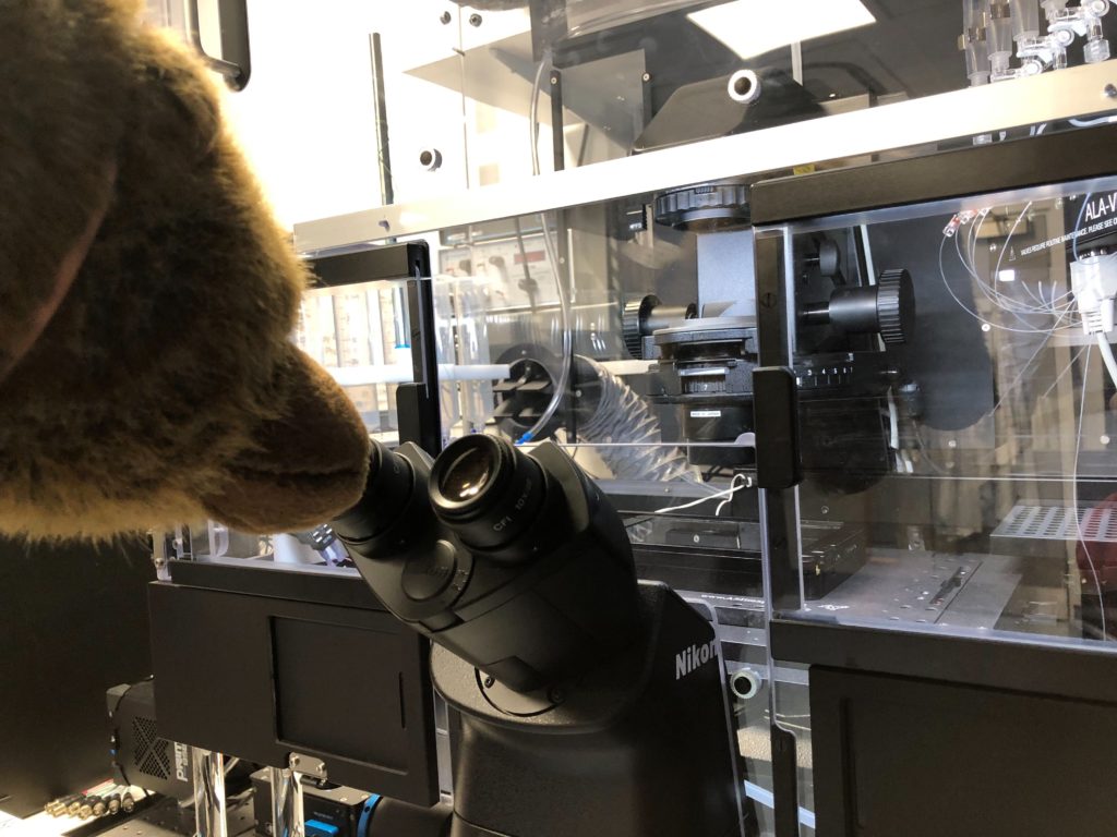
Up-close with a microscope!
Thanks to Hector and Joao for inviting us to COMPARE and showing us their facilities!
Look out for our next researcher soon!